Definition
Uveitis is swelling and irritation of the uvea, the middle layer of the eye. The uvea provides most of the blood supply to the retina.
Overview, Causes, & Risk Factors
Uveitis can be caused by autoimmune disorders such as rheumatoid arthritis or ankylosing spondylitis, infection, or exposure to toxins. However, in many cases the cause is unknown.
The most common form of uveitis is anterior uveitis, which involves inflammation in the front part of the eye. It is often called iritis because it is usually only effects the iris, the colored part of the eye. The inflammation may be associated with autoimmune diseases, but most cases occur in healthy people. The disorder may affect only one eye. It is most common in young and middle-aged people.
Posterior uveitis affects the back part of the uvea, and involves primarily the choroid, a layer of blood vessels and connective tissue in the middle part of the eye. This type of uveitis is called choroiditis. If the retina is also involved it is called chorioretinitis. You may develop this condition if you have had a body-wide (systemic) infection or if you have an autoimmune disease.
Another form of uveitis is pars planitis. This inflammation affects the narrow area between the colored part of the eye (iris) and the choroid. Pars planitis usually occurs in young men and is generally not associated with any other disease. However, some evidence suggests it may be linked to Crohn’s disease and possibly multiple sclerosis.
Uveitis can be associated with any of the following:
- AIDS
- Ankylosing spondylitis
- Behcet syndrome
- CMV retinitis
- Herpes zoster infection
- Histoplasmosis
- Injury
- Kawasaki disease
- Psoriasis
- Reactive arthritis
- Rheumatoid arthritis
- Sarcoidosis
- Syphilis
- Toxoplasmosis
- Tuberculosis
- Ulcerative colitis
Pictures & Images
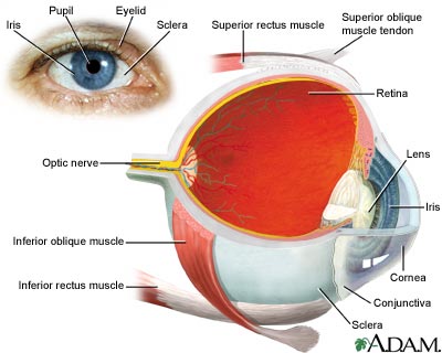
Eye
The eye is the organ of sight, a nearly spherical hollow globe filled with fluids (humors). The outer layer or tunic (sclera, or white, and cornea) is fibrous and protective. The middle tunic layer (choroid, ciliary body and the iris) is vascular. The innermost layer (the retina) is nervous or sensory. The fluids in the eye are divided by the lens into the vitreous humor (behind the lens) and the aqueous humor (in front of the lens). The lens itself is flexible and suspended by ligaments which allow it to change shape to focus light on the retina, which is composed of sensory neurons.
Visual field test
Central and peripheral vision is tested by using visual field tests. Changes may indicate eye diseases, such as glaucoma or retinitis.
Review Date : 7/28/2008
Reviewed By : Manju Subramanian, MD, Assistant Professor in Ophthalmology, Vitreoretinal Disease and Surgery, Boston University Eye Associates, Boston, MA. Review provided by VeriMed Healthcare Network. Also reviewed by David Zieve, MD, MHA, Medical Director, A.D.A.M., Inc.
![]()
