Definition
Tinea versicolor is a long-term (chronic) fungal infection of the skin.
Overview, Causes, & Risk Factors
Tinea versicolor is relatively common. It is caused by the fungus Pityrosporum ovale, a type of yeast that is normally found on human skin. It only causes problems under certain circumstances.
The condition is most common in adolescent and young adult males. It typically occurs in hot climates.
Pictures & Images
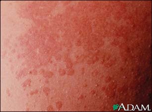
Tinea versicolor – close-up
Tinea versicolor is a superficial fungal infection common in adolescent and young adult males. This close-up view demonstrates the typical pattern of the rash.
Tinea versicolor – shoulders
Tinea versicolor is a superficial fungal infection common in adolescent and young adult males. Frequent sites of infection include the neck, upper chest, and axilla (arm pit). The rash may range from yellow to golden brown in color. Mild itching is also associated with this infection. This photograph demonstrates fairly extensive involvement.
Tinea versicolor – close-up
This is a fungal infection of the skin known as tinea versicolor, and is common in adolescent and young adult males. Besides the rash, there may be mild itching. Frequent sites of infection include the neck, upper chest, and arm pit (axilla). The rash may be white to yellowish to golden brown in color. A tan can accentuate the difference in skin color.
Tinea versicolor on the back Tinea versicolor is an infection caused by a fungus that is common in adolescent and young adult males. Besides the rash, seen here on the back, there may be mild itching. Frequent sites of infection include the neck, upper chest, and arm pit (axilla). The rash may be white (as seen here) to yellowish to golden brown in color. A tan can accentuate the difference in skin color.

Tinea versicolor is caused by the organism Pityrosporum ovale. It occurs most often in young adults. Wood’s lamp examination revelas pale yellow-green fluorescence. KOH prep reveals “spaghetti and meatballs” with hyphae and spores. Skin lesions are sharply marginated macules, either hyper or hypopigmented, covered with fine scale. Small discrete lesions may eventually coalesce to cover large areas of the trunk.
-
Tinea versicolor: Overview, Causes
-
Tinea versicolor: Symptoms & Signs, Diagnosis & Tests
-
Tinea versicolor: Treatment
Review Date : 10/28/2008
Reviewed By : Michael Lehrer, MD, Department of Dermatology, University of Pennsylvania Medical Center, Philadelphia, PA. Review provided by VeriMed Healthcare Network. Also reviewed by David Zieve, MD, MHA, Medical Director, A.D.A.M., Inc.
![]()
