Definition
Tinea corporis is a skin infection due to fungi.
See also:
- Tinea capitis
- Tinea cruris (jock itch)
- Tinea pedis (athlete’s foot)
Overview, Causes, & Risk Factors
Tinea corporis (often called ringworm of the body) is a common skin disorder, especially among children. However, it may occur in people of all ages. It is caused by mold-like fungi called dermatophytes.
Fungi thrive in warm, moist areas. The following raise your risk for a fungal infection:
- Long-term wetness of the skin (such as from sweating)
- Minor skin and nail injuries
- Poor hygiene
Tinea corporis is contagious. You can catch the condition if you come into direct contact with someone who is infected, or if you touch contaminated items such as:
- Clothing
- Combs
- Pool surfaces
- Shower floors and walls
The fungi can also be spread by pets (cats are common carriers).
Pictures & Images
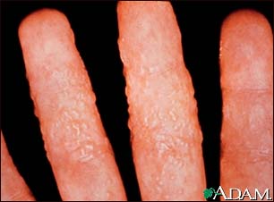
Dermatitis, reaction to tinea
 This picture shows a skin inflammation of the fingers with multiple blisters (vesicles) caused by an allergic reaction to a fungal infection (tinea corporis). (Image courtesy of the Centers for Disease Control and Prevention.)
Tinea versicolor – close-up
 This child’s leg shows a classical-appearing ringworm lesion with central clearing and a slightly raised red border.This is a fungal infection of the skin known as tinea versicolor, and is common in adolescent and young adult males. Besides the rash, there may be mild itching. Frequent sites of infection include the neck, upper chest, and arm pit (axilla). The rash may be white to yellowish to golden brown in color. A tan can accentuate the difference in skin color.
Tinea versicolor on the back
 Tinea versicolor is an infection caused by a fungus that is common in adolescent and young adult males. Besides the rash, seen here on the back, there may be mild itching. Frequent sites of infection include the neck, upper chest, and arm pit (axilla). The rash may be white (as seen here) to yellowish to golden brown in color. A tan can accentuate the difference in skin color.
Ringworm, tinea corporis on an infant’s leg
 This child’s leg shows a classical-appearing ringworm lesion with central clearing and a slightly raised red border.
Ringworm, tinea manuum on the finger
 This is a picture of ringworm, tinea manum, on the finger. This fungal infection is inflamed and scaly.
Tinea versicolor – close-up

Tinea versicolor is a superficial fungal infection common in adolescent and young adult males. This close-up view demonstrates the typical pattern of the rash.
Tinea versicolor – shoulders

Tinea versicolor is a superficial fungal infection common in adolescent and young adult males. Frequent sites of infection include the neck, upper chest, and axilla (arm pit). The rash may range from yellow to golden brown in color. Mild itching is also associated with this infection. This photograph demonstrates fairly extensive involvement.
Ringworm, tinea on the hand and leg

This is a picture of ringworm (tinea) on the hand and leg. Tinea is a fungal infection of the skin. Ringworm is not seen as frequently in adults as in children, but when conditions are conducive to growth, the fungus can flourish.
Ringworm, tinea corporis on the leg
 Ringworm is a fungal infection of the skin. It usually produces a ring-shaped lesion which appears to clear in the center. The edges of the lesion may be slightly raised and often itch. Central clearing can be seen in some of the infected areas on the leg of this person.
Granuloma, fungal (Majocchi’s)
 This condition of fungal granuloma has produced a large, red (erythematous) patch (plaque) with a prominent border, within which are scattered blisters (pustules) indicating deeper involvement of hair follicles (Majocchi’s granuloma). This infection was caused by Trichophyton rubrum.
Granuloma, fungal (Majocchi’s)

This is a picture of a fungal granuloma, a large, red (erythematous) patch (plaque) with a prominent border. Within the borders of the lesion are scattered blisters (pustules) that indicate deeper involvement of hair follicles (Majocchi’s granuloma). This dermatophyte infection was caused by Trichophyton rubrum.
Tinea corporis – ear

Tinea coporis of the ear is shown here. The area is erythematous with peripheral scale. There is some erosion and fissuring, likely from the inflammation incited by the dermatophyte. Topical antifungal cream may be curative.
-
Tinea corporis: Overview, Causes
-
Tinea corporis: Symptoms & Signs, Diagnosis & Tests
-
Tinea corporis: Treatment
Review Date : 10/3/2008
Reviewed By : Kevin Berman, MD, PhD, Atlanta Center for Dermatologic Disease, Atlanta, GA. Review provided by VeriMed Healthcare Network. Also reviewed by David Zieve, MD, MHA, Medical Director, A.D.A.M., Inc.
![]()
