Alternate Names : Miliary tuberculosis, Tuberculosis – disseminated, Extrapulmonary tuberculosis
Definition
Disseminated tuberculosis (TB) is a contagious bacterial infection that has spread from the lungs to other parts of the body through the blood or lymph system.
See also: Tuberculosis – pulmonary
Overview, Causes, & Risk Factors
Tuberculosis (TB) infection can develop after inhaling droplets sprayed into the air from a cough or sneeze by someone infected with the Mycobacterium tuberculosis bacteria. Small areas of infection, called granulomas (granular tumors), develop in the lungs.
The usual site of tuberculosis is the lungs, but other organs can be involved. In the U.S., most people with primary tuberculous get better and have no further evidence of disease. Disseminated disease develops in the small number of infected people whose immune systems do not successfully contain the primary infection.
Disseminated disease can occur within weeks after the primary infection, or may lie dormant for years before causing illness. Infants, the elderly, those infected with HIV. and those who take immune-suppressing medications are at higher risk for the disease worsening, because of their weaker immune systems.
In disseminated disease, organs and tissues affected can include:
- Bones and joints
- Bronchus
- Cervical lymph nodes
- Eye
- Larynx (voice box)
- Lining of the abdominal cavity (peritoneum)
- Lining of the brain and spinal cord (meninges)
- Lining of the heart (pericardium)
- Organs of the male or female urinary and reproductive systems
- Skin
- Small bowel
- Stomach
The risk of catching TB increases when you are in contact with people who have the disease, if you live in crowded or unsanitary conditions, and if you have poor nutrition.
Recently, TB has been seen more often in the U.S. Factors that may be causing this increase are tuberculosis infections in people with AIDS and HIV, and increasing numbers of homeless people.
Another matter of concern is the development of drug-resistant strains of TB. Incomplete treatment of TB infections (such as not taking medications for the prescribed length of time) can contribute to the development of drug-resistant strains of bacteria.
About half of AIDS patients with a CD4 count less than 200 who develop TB will have disseminated disease (not localized disease, as in pulmonary tuberculosis).
Pictures & Images
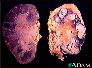
Tuberculosis in the kidney
 Kidneys can be damaged by tuberculosis. Tuberculosis generally affects the lungs, but may cause infection in many other organs in the body. (Image courtesy of the Centers for Disease Control and Prevention.)
Tuberculosis in the lung

Tuberculosis is caused by a group of organisms Mycobacterium tuberculosis, M. bovis, M. africanum and a few other rarer subtypes. Tuberculosis usually appears as a lung (pulmonary) infection. However, it may infect other organs in the body. Recently, antibiotic-resistant strains of tuberculosis have appeared. With increasing numbers of immunocompromised individuals with AIDS, and homeless people without medical care, tuberculosis is seen more frequently today. (Image courtesy of the Centers for Disease Control and Prevention.)
Coal worker’s lungs – chest x-ray

This chest x-ray shows coal worker’s lungs. There are diffuse, small, light areas on both sides (1 to 3 mm) in all parts of the lungs. Diseases that may result in an x-ray like this include: simple coal workers pneumoconiosis (CWP) – stage I, simple silicosis, miliary tuberculosis, histiocytosis X (eosinophilic granuloma), and other diffuse infiltrate pulmonary diseases.
Coal workers pneumoconiosis – stage II

This chest x-ray shows stage II coal worker’s pneumoconiosis (CWP). There are diffuse, small light areas on both sides of the lungs. Other diseases�that may explain these x-ray findings include simple silicosis, disseminated tuberculosis, metastatic lung cancer, and other diffuse, infiltrative pulmonary diseases.
Coal workers pneumoconiosis, complicated

This picture shows complicated coal workers pneumoconiosis. There are diffuse, small, light areas (3 to 5 mm) in all areas on both sides of the lungs. There are large light areas which run together with poorly defined borders in the upper areas on both sides of the lungs. Diseases which may explain these X-ray findings include complicated coal workers pneumoconiosis (CWP), silico-tuberculosis, disseminated tuberculosis, metastatic lung cancer, and other diffuse infiltrative pulmonary diseases.
Tuberculosis, advanced – chest x-rays

Tuberculosis is an infectious disease that causes inflammation, the formation of tubercules and other growths within tissue, and can cause tissue death. These chest x-rays show advanced pulmonary tuberculosis. There are multiple light areas (opacities) of varying size that run together (coalesce). Arrows indicate the location of cavities within these light areas. The x-ray on the left clearly shows that the opacities are located in the upper area of the lungs toward the back. The appearance is typical for chronic pulmonary tuberculosis but may also occur with chronic pulmonary histiocytosis and chronic pulmonary coccidioidomycosis. Pulmonary tuberculosis is making a comeback with new resistant strains that are difficult to treat. Pulmonary tuberculosis is the most common form of the disease, but other organs can be infected.
Sarcoid, stage II – chest x-ray

Sarcoid causes damage to the lung tissue that heals by scarring. The film shows a diffuse milky and granular appearance in the normally dark lung areas. This individual has marked decrease in lung function.
Miliary tuberculosis

Miliary tuberculosis is characterized by a chronic, contagious bacterial infection caused by Mycobacterium tuberculosis that has spread to other organs of the body by the blood or lymph system.
Erythema multiforme, circular lesions – hands

Erythema multiforme lesions are circular and may appear in concentric rings (often called target lesions). Target lesions may also be associated with other medical conditions such as herpes infection, streptococcal infection, tuberculosis (TB), or as a reaction to chemicals or medications.
Erythema nodosum associated with sarcoidosis

This picture shows reddish-purple, hard (indurated), painful nodules (Erythema nodosum) that occur most commonly on the shins. These lesions may be anywhere on the body and may be associated with tuberculosis (TB), sarcoidosis, coccidioidomycosis, systemic lupus erythematosis (SLE), fungal infections, or in response to medications.
Circulatory system

Blood used by the body is brought back to the heart and lungs by the veins of the body. Once the blood has gathered more oxygen from the lungs, it is pumped back out to the body through the arteries.
-
Disseminated tuberculosis : Overview, Causes, & Risk Factors
-
Disseminated tuberculosis : Symptoms & Signs, Diagnosis & Tests
-
Disseminated tuberculosis : Treatment


Review Date : 12/1/2009
Reviewed By : David C. Dugdale, III, MD, Professor of Medicine, Division of General Medicine, Department of Medicine, University of Washington School of Medicine; Jatin M. Vyas, MD, PhD, Assistant Professor in Medicine, Harvard Medical School, Assistant in Medicine, Division of Infectious Disease, Department of Medicine, Massachusetts General Hospital. Also reviewed by David Zieve, MD, MHA, Medical Director, A.D.A.M., Inc.
The information provided herein should not be used during any medical emergency or for the diagnosis or treatment of any medical condition. A licensed medical professional should be consulted for diagnosis and treatment of any and all medical conditions. Call 911 for all medical emergencies. Links to other sites are provided for information only — they do not constitute endorsements of those other sites. © 1997- 2010 A.D.A.M., Inc. Any duplication or distribution of the information contained herein is strictly prohibited.
