Alternate Names : Rodent ulcer, Skin cancer – basal cell, Cancer – skin – basal cell
Definition
Basal cell carcinoma is a slow-growing form of skin cancer.
See also:
- Squamous cell skin cancer
- Melanoma
Overview, Causes, & Risk Factors
Skin cancer is divided into two major groups: nonmelanoma and melanoma. Basal cell carcinoma is a type of nonmelanoma skin cancer, and is the most common form of cancer in the United States. According to the American Cancer Society, 75% of all skin cancers are basal cell carcinomas.
Basal cell carcinoma starts in the top layer of the skin called the epidermis. It grows slowly and is painless. A new skin growth that bleeds easily or does not heal well may suggest basal cell carcinoma. The majority of these cancers occur on areas of skin that are regularly exposed to sunlight or other ultraviolet radiation. They may also appear on the scalp. Basal cell skin cancer used to be more common in people over age 40, but is now often diagnosed in younger people.
Your risk for basal cell skin cancer is higher if you have:
- Light-colored skin
- Blue or green eyes
- Blond or red hair
- Overexposure to x-rays or other forms of radiation
Basal cell skin cancer almost never spreads. But, if left untreated, it may grow into surrounding areas and nearby tissues and bone.
Pictures & Images
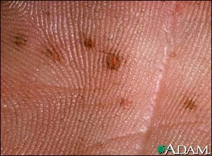
Basal cell nevus syndrome – close-up of palm
Basal cell nevus syndrome is an inherited disorder characterized by wide-set eyes, saddle nose, frontal bossing (prominent forehead), prognathism (prominent chin), numerous basal cell carcinomas, and skeletal abnormalities. Skin manifestations include pits in the palms and soles, and numerous basal cell carcinomas. This picture is a close-up of the pits found in the palm of an individual with basal cell nevus syndrome.
Skin cancer, basal cell carcinoma – nose

The typical basal cell skin cancer appears as a small, pearly, dome-shaped nodule with small visible blood vessels (telangiectasias).
Skin cancer, basal cell carcinoma – pigmented

This skin cancer appears as a 2 to 3 centimeter skin spot. The tissue has become destroyed (forming an atrophic plaque). There is a brownish color because of increased skin pigment (hyperpigmentation) and a slightly elevated, rolled, pearl-colored margin. This growth is located along the hair line.
Skin cancer, basal cell carcinoma – behind ear

This skin cancer appears as a 1 to 1.5 centimeter flesh-colored nodule with a central depression and a raised, pearly border. Small blood vessels are visible (telangiectatic).
Skin cancer, basal cell carcinoma – spreading

This skin cancer, a basal cell carcinoma, is 5 to 6 centimeters across, red (erythematous), with well defined (demarcated) borders and sprinkled brown pigment along the margins. This cancer is located on the person’s back.
Basal cell nevus syndrome – plantar pits

Basal cell nevus syndrome is an inherited disorder characterized by wide-set eyes, saddle nose, frontal bossing (prominent forehead), prognathism (prominent chin), and skeletal abnormalities. Skin manifestations include pits in the palms and soles, and numerous basal cell carcinomas (skin cancers). This picture is a close-up of the pits found on the sole of the foot of an individual with basal cell nevus syndrome.
Basal cell nevus syndrome – face and hand

Basal cell nevus syndrome is an inherited disorder characterized by wide-set eyes, saddle nose, frontal bossing (prominent forehead), prognathism (prominent chin), numerous basal cell carcinomas (a type of skin cancer), and skeletal abnormalities. This individual has multiple flesh-colored, dome-shaped bumps on the face which are basal cell cancers, and palmar pits.
Multiple basal cell cancer due to x-ray therapy for acne

Basal cell carcinomas are more prevalent on sun or radiation exposed areas of skin. Here the typical lesion with raised, rolled, pearly borders with ulcerated center is seen on the back of a person previously irradiated for acne.
Basal cell carcinoma – nose

A basal cell carcinoma near the tip of the nose is shown here. This lesion has a pearly, slightly raised border, and would likely bleed easily if traumatized.
Basal Cell Carcinoma – face

This flesh-colored, verrucal, pearly, smooth, non-scaling papule is a basal cell carcinoma. It is occurring in a common, sun-exposed area of the face, on the forehead.
Basal Cell Carcinoma – close-up

This basal cell carcinoma exhibits a characteristic of this type of lesion-telangiectasia. The lesion is also pearly, and smooth, with a slight central depression.
Basal cell carcinoma – close-up

This basal cell carcinoma appears as a multicolored flat lesion, with a periphery that has ulcerated and bled. Telangiectasia are present.
Basal cell cancer

Basal cell cancer is a malignant skin tumor involving cancerous changes of basal skin cells. Basal cell skin cancers usually occur on areas of skin that are regularly exposed to sunlight or other ultraviolet radiation. Once a suspicious lesion is found, a biopsy is needed to prove the diagnosis of basal cell carcinoma. Treatment varies depending on the size, depth, and location of the cancer. Early treatment by a dermatologist may result in a cure rate of more than 95%, but regular examination by a health care provider is required to watch for new sites of basal cell cancer.
-
Basal cell carcinoma : Overview, Causes, & Risk Factors
-
Basal cell carcinoma : Symptoms & Signs, Diagnosis & Tests
-
Basal cell carcinoma : Treatment


Review Date : 2/5/2008
Reviewed By : Kevin Berman, MD, PhD, Associate, Atlanta Center for Dermatologic Disease, Atlanta, GA. Review provided by VeriMed Healthcare Network. Also reviewed by David Zieve, MD, MHA, Medical Director, A.D.A.M., Inc.

